The Nuclear Camera (or otherwise known as a gamma camera) uses special imaging techniques called planar dynamic or single-photon emission-computed tomography (SPECT) to detect radioactive energy that is emitted from the patient’s body and convert it into an image. The gamma camera itself does not emit any radiation. It is composed of radiation detectors, called gamma camera heads, which are encased in metal and plastic and most often shaped like a box, attached to a round circular donut shaped gantry.
The radioactive energy emitting from the patient’s body is the result of the introduction of a radioactive material called a radiopharmaceutical or radiotracer into the body. This material is either injected into the bloodstream, swallowed or inhaled as a gas. The material accumulates in the area of your body under examination, where it gives off a small amount of energy in the form of gamma rays. The gamma camera detects this energy and, with the help of a computer, create pictures that offer details on the structure and function of organs and tissues.
During the Exam
Depending on your type of nuclear medicine exam, the radiotracer is injected intravenously, swallowed or inhaled as a gas.
It can take anywhere from several seconds to a number of days for the radiotracer to travel through your body and accumulate in the area under study. As a result, imaging may be done immediately, a few hours later, or even days after you receive the radioactive material.
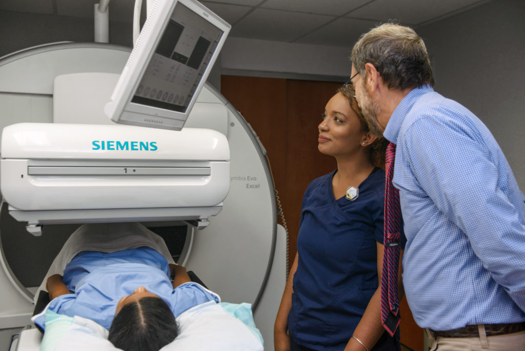
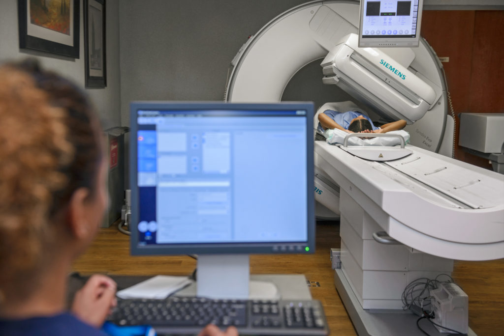
When it is time for the imaging to begin, you will be asked to lie on an examination table which slides in between two gamma camera heads that are positioned above and below you. The camera or scanner will take a series of images. The camera may rotate around you or it may stay in one position and you may be asked to change positions in between images. While the camera is taking pictures, you will need to remain still for brief periods of time. In some cases, the camera may move very close to your body. This is necessary to obtain the best quality images. If you are claustrophobic, you should inform the technologist before your exam begins.
The length of time for nuclear medicine procedures varies greatly, depending on the type of exam. Actual scanning time for Nuclear Imaging exams can take from 20 minutes to several hours and may be conducted over multiple days.
When the examination is complete, you may be asked to wait until the technologist checks the images in case more images are needed. Sometimes, more images are obtained to clarify or better visualize certain areas or structures. The need for more images does not necessarily mean there was a problem with the exam or that something abnormal was found.
Preparation
You may wear a gown during the exam or be allowed to wear your own clothing.
Women should always tell their doctor and technologist if there is any possibility that they are pregnant or if they are breastfeeding.
Tell the doctor and the technologist performing your exam about any medications you are taking, including vitamins and herbal supplements. List any allergies, recent illnesses and other medical conditions.
Leave jewelry and other metallic accessories (including, buttons and zippers) at home or remove them prior to the exam. Such objects may interfere with the procedure.
Inform the doctor or the technologist if you work with or have small children at home. Finally, inform the doctor or technologist if you have any travel plans soon after the study so you can be provided with the proper documentation.
Our technologist and an appointment specialist will contact you with specific instructions based on the type of scan you are receiving in advance of your exam.
Bone scans aren’t usually performed on pregnant women or nursing mothers because of concerns about radiation exposure to the baby. Tell us if you are pregnant (or think you might be pregnant) or if you are nursing.