Magnetic Resonance Imaging (MRI) uses a powerful magnetic field, radio waves and a computer to produce detailed pictures of the body’s internal structures that are clearer, more detailed and more likely in some instances to identify and accurately characterize disease than other imaging methods. There are no known risks from an MRI scan as it is non-invasive and does not expose you to any radiation. As a result, a patient may have multiple MRI scans without being concerned about radiation exposure.
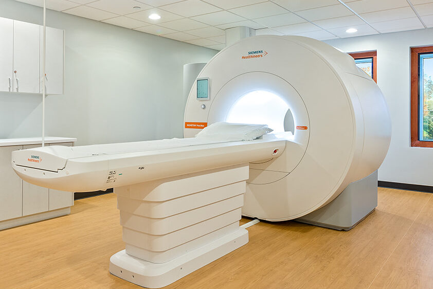
MAGNETOM Free.Max Wide-Open MRI

3T MRI
Instead of radiation, MRI uses magnetic fields and radio frequency to momentarily move the molecules in your body out of their normal alignment or their normal spinning pattern without you even physically feeling it. As the molecules return to their natural positions, the MRI machine records that activity and uses the information to create detailed images of the organs, tissues and other structures inside your body. The radiologist then analyzes the images either layer by layer or as 3D structures.
The typical (closed) MRI machine looks like a narrow tube that has both ends open. Our MAGNETOM Free.Max MRI machine has the largest bore on the market today, providing our patients a wide-open alternative to the closed machines. For patients who experience claustrophobia or whose bodies do not comfortably fit into a narrower tube the MAGNETOM Free.Max is a phenomenal solution. Not only can the MAGNETOM Free.Max accommodate patients in excess of 500lbs, its technology harnesses the power of AI to deliver impressive imaging. UDMI offers both closed (3T) MRI as well as a the MAGENETOM Free.Max Wide-Open MRI. The radiologist along with you and the MRI technologist will make the determination which machine is more appropriate for your exam. Learn more about the 3T Magnetic Resonance Imaging (MRI) machine and its capabilities.
During the Exam
During the MRI exam, the patient is asked to rest motionless on a padded table. A typical exam may last as little as 10 minutes, while more involved studies can last as long as 40 minutes. The anatomic area of interest will be positioned in the center of the field. During the scan the patient does not feel a thing but will hear faint hums and the thumping of radio waves. To help make the experience a more pleasant one, we provide headphones for the patient to listen to music during the exam.
MAGNETOM Free.Max with patient

3T MRI with patient

Once the patient is positioned correctly, the technologist will leave the room to operate the MRI machine from a workstation in a control room. During the MRI exam, the technologist and patient can communicate at any time using an intercom. The technologist watches the patient during the study and monitors the progress of the image during the study.
Depending on the part of the body being scanned different contrast materials may be used. Contrast materials are administered via intravenous administration. Contrast is a liquid that enhances the images of the organs and/or blood vessels. It is safe with minimal contraindications. The contrast is cleared by the kidneys and is typically out of the patient’s body in 3-4 hours. The decision whether or not contrast material will be of value to your study will be made by the our radiologists and your referring physician. Learn more about our 3T MRI
Preparation
Patients can continue to take all medication and follow their regular diet unless instructed otherwise. Prior to entering the scan room, the patient will be instructed to remove all jewelry, hairclips, watches, coins, keys and any other metal objects from their body. Credit cards and ATM cards must not be brought into the scan room, as the magnet will damage the magnetic strip on the cards.
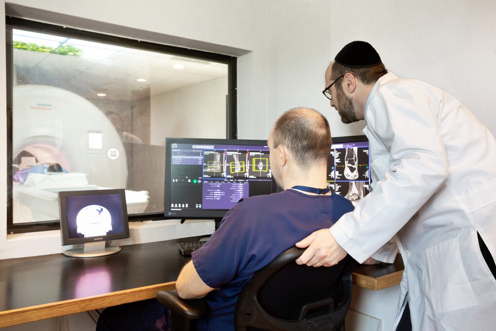
3T MRI Tech View
When making your appointment tell us if you:
- Have a pacemaker
- Are pregnant, or suspect that you may be
- Have aneurysm clips
- Have had heart or brain surgery
- Have had orthopedic spine surgery where metallic hardware was placed
- Have had metal fragments in your eyes
- Have shrapnel in your body
- Suffer from claustrophobia
- Weigh 300 lbs. or more
If your MRI exam requires the use of intravenous contrast, we may need bloodwork from your referring physician to ensure it is safe for you to receive the injection.