Vascular Imaging uses Ultrasound, Computed Tomography (CT), and Magnetic Resonance Imaging (MRI) to evaluate the body’s circulatory system to help identify blockages in the arteries and veins and detect blood clots.
Ultrasound is particularly useful to evaluate the body’s vessels in real time. CT and MRI are useful in visualizing vessels that either cannot be seen by ultrasound or require additional evaluation.
UDMI commonly performs the following vascular imaging exams:
Ultrasound
- Carotid
- Transcranial Doppler (TCD)
- Aorta
- Upper / Lower Extremity: arterial or venous
- Venous Duplex: For reflux or deep vein thrombosis (DVT)
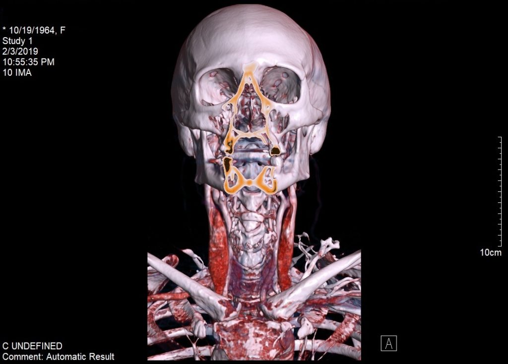
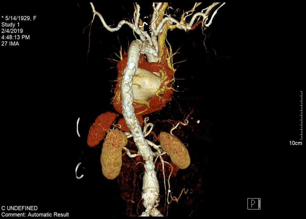
CTA / MRA
- Head
- Neck
- Chest Abdomen
- Pelvis
- Upper / Lower Extremity
- CTA / MRA for pulmonary embolism
- CTA / MRA for aneurysm (chest, abdomen, pelvis upper / lower extremity)