Pelvic Ultrasound

Ultrasound uses sound waves to create pictures of the uterus and ovaries. It is safe and painless. Ultrasound of the pelvis is either performed by placing the probe on the patient’s lower abdomen (transabdominal) or in the vagina (also referred to as endovaginal Ultrasound). An endovaginal Ultrasound resembles a pelvic exam performed by your gynecologist. A thin covered wand or probe is placed into the vagina and the examiner directs the probe toward the uterus and ovaries. It is not unusual for women to have both transabdominal and endovaginal Ultrasound on the same visit, as the exams provide different information to your physician.
Ultrasound is also used to guide procedures such as a sonohysterogram, which allows for a close look of the lining of the uterus.
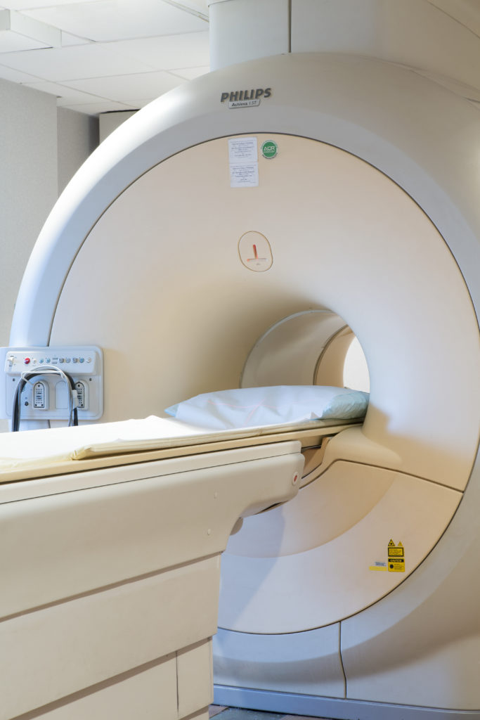
Pelvic MRI
Magnetic Resonance Imaging (MRI) of the pelvis uses a powerful magnetic field, radio waves and a computer to create detailed pictures of the uterus, ovaries, and nearby structures such as the bladder. It is often used in conjunction with pelvic sonography to evaluate problems such as an enlarged uterus, ovarian masses, and lymphovascular abnormalities. In most cases, the MRI is performed with intravenous contrast which enhances the visibility of soft tissue differentiation and helps better define any abnormalities encountered.
Hysterosalpingogram (HSG)
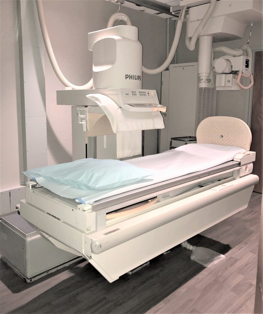
Hysterosalpingogram (HSG) is a procedure that uses an x-ray and contrast to look at the fallopian tubes and uterus of a woman who is having difficulty becoming pregnant. It is also used to investigate the cause of miscarriages resulting from abnormalities within the uterus and to determine the presence and severity of tumor masses, adhesions and uterine fibroids.